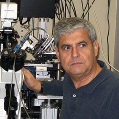
Julio Vergara
School of Medicine
Los Angeles, CA 90095 School of Medicine
Los Angeles, CA 90095
Office Phone Number:
310-825-9307
Affiliations
Research Interests
Biography
Biophysics of the Excitation-Contraction Coupling process in Skeletal Muscle. Research in our laboratory aims to unveil the detailed mechanisms of excitation-contraction (EC) coupling in vertebrate skeletal muscle and their pathological alterations in diseases. To this end, the laboratory uses a combination of advanced optical and electrophysiological approaches. The group has shown, with a localized detection method and pulse laser imaging that one of the earliest manifestations of the EC coupling process in skeletal muscle is a massive local increase of the intracellular Ca2+ concentration in microscopic regions of the muscle fibers where the sarcoplasmic reticulum (SR) apposes the T-tubule (triadic junction). It has also been recently found that these Ca2+ microdomains in mammalian muscle fibers are doubled peaked, as expected from the existence of two triadic junctions per sarcomere. The large, rapidly dissipating, Ca2+ gradients are not restricted to action potential-evoked Ca2+ release in muscle fibers; they also occur as a triggering mechanism in synaptic transmission and other neurosecretory processes. Novel approaches in imaging, flash-photolysis, and de-novo expression of recombinant membrane proteins are currently under development. These methodologies will allow the group to obtain new insights on the role of the membrane potential in initiating the cascade of events that lead to EC coupling in skeletal muscle fibers.
Publications
A selected list of publications:
- DiFranco Marino, Vergara Julio L The Na conductance in the sarcolemma and the transverse tubular system membranes of mammalian skeletal muscle fibers The Journal of general physiology, 2011; 138(4): 393-419.
- Ermolova Natalia, Kudryashova Elena, DiFranco Marino, Vergara Julio, Kramerova Irina, Spencer Melissa J Pathogenity of some limb girdle muscular dystrophy mutations can result from reduced anchorage to myofibrils and altered stability of calpain 3 Human molecular genetics, 2011; 20(17): 3331-45.
- DiFranco Marino, Tran Philip, Quiñonez Marbella, Vergara Julio L Functional expression of transgenic 1sDHPR channels in adult mammalian skeletal muscle fibres The Journal of physiology, 2011; 589(Pt 6): 1421-42.
- DiFranco Marino, Herrera Alvaro, Vergara Julio L Chloride currents from the transverse tubular system in adult mammalian skeletal muscle fibers The Journal of general physiology, 2011; 137(1): 21-41.
- Capote Joana, DiFranco Marino, Vergara Julio L Excitation-contraction coupling alterations in mdx and utrophin/dystrophin double knockout mice: a comparative study American journal of physiology. Cell physiology, 2010; 298(5): C1077-86.
- DiFranco Marino, Quinonez Marbella, Capote Joana, Vergara Julio DNA transfection of mammalian skeletal muscles using in vivo electroporation Journal of visualized experiments : JoVE, 2009; 298(32): .
- DiFranco Marino, Capote Joana, Quiñonez Marbella, Vergara Julio L Voltage-dependent dynamic FRET signals from the transverse tubules in mammalian skeletal muscle fibers J. Gen. Physiol, 2007; 130(6): 581-600.
- Faas Guido C, Schwaller Beat, Vergara Julio L, Mody Istvan Resolving the fast kinetics of cooperative binding: Ca2+ buffering by calretinin PLoS Biol, 2007; 5(11): e311.
- Gómez José, Neco Patricia, DiFranco Marino, Vergara Julio L Calcium release domains in mammalian skeletal muscle studied with two-photon imaging and spot detection techniques J. Gen. Physiol, 2006; 127(6): 623-37.
- DiFranco Marino, Neco Patricia, Capote Joana, Meera Pratap, Vergara Julio L Quantitative evaluation of mammalian skeletal muscle as a heterologous protein expression system Protein Expr. Purif, 2006; 47(1): 281-8.
- Woods Christopher E, Novo David, DiFranco Marino, Capote Joana, Vergara Julio L Propagation in the transverse tubular system and voltage dependence of calcium release in normal and mdx mouse muscle fibres J. Physiol. (Lond.), 2005; 568(Pt 3): 867-80.
- Faas Guido C, Karacs Kinga, Vergara Julio L, Mody Istvan Kinetic properties of DM-nitrophen binding to calcium and magnesium Biophys. J, 2005; 88(6): 4421-33.
- Woods Christopher E, Novo David, DiFranco Marino, Vergara Julio L The action potential-evoked sarcoplasmic reticulum calcium release is impaired in mdx mouse muscle fibres J. Physiol. (Lond.), 2004; 557(Pt 1): 59-75.
- Novo David, DiFranco Marino, Vergara Julio L Comparison between the predictions of diffusion-reaction models and localized Ca2+ transients in amphibian skeletal muscle fibers Biophys. J, 2003; 85(2): 1080-97.
